Doctors perform complex, rare surgery on grape-size heart of foetus still in mother’s womb
Sign up now: Get insights on Asia's fast-moving developments

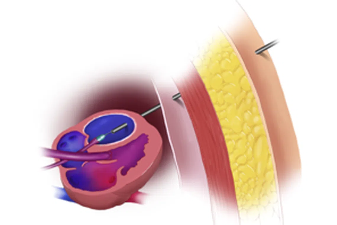
This procedure, called balloon dilation of the aortic valve, is a rare and very delicate but quick procedure.
PHOTO: TEXAS CHILDREN'S HOSPITAL
A team of elite doctors in India has performed what amounts to a medical jiu-jitsu: They stuck a long needle through the abdomen of a woman to enlarge a valve in the grape-size heart of the foetus inside her.
This procedure is called “balloon dilation of the aortic valve”, a rare and very delicate procedure that is also an amazingly quick one. The surgeons’ feat took 90 seconds.
The surgery was performed at the All India Institute of Medical Sciences (AIIMS) in New Delhi.
It was such a landmark medical feat that India’s Prime Minister Narendra Modi tweeted: “Proud of India’s doctors for their dexterity and innovation.”
Union Health Minister Mansukh Mandaviya also took to Twitter to congratulate the AIIMS cardiologists and foetal medicine specialists involved.
The Times of India reported that the 28-year-old mother has had three miscarriages already, so she was determined to keep the foetus she is now carrying.
But the foetus had a severely narrowed aortic valve, a condition known as “aortic stenosis”, a complex congenital heart defect that occurs when the left side of the heart does not form properly.
Without intervention, the left ventricle in foetuses with aortic stenosis often stops growing, which will require immediate surgery after birth.
The Texas Children’s Hospital said on its website that most children born with aortic stenosis will require at least three cardiac “plumbing” operations before the age of six, and most will need a heart transplant later in life.
The doctors at AIIMS determined that performing balloon dilation while the foetus was in its mother’s womb “may improve the outlook for the baby after birth and lead to near normal development”.
Balloon dilation is an ultrasound-guidance procedure.
The surgeon inserts a special needle through the mother’s abdomen and uterus, and into the baby’s heart.
Once in position, a wire with an attached balloon is threaded through the needle and across the narrowed aortic valve.
The tiny balloon is then gently inflated, enlarging the opening of the aortic valve and increasing blood flow into the left ventricle. It is then deflated, and the balloon, wire and needle are removed.
“Such a procedure is very challenging, as it can risk even the life of a foetus. It has to be done very precisely... and then it has to be done very quickly because you risk puncturing the major heart chamber,” one of the senior AIIMS doctors involved in the surgery said.
“If something does go wrong,” he said, “the baby will die. It has to be very quick: shoot and dilate and come out.”
He said the mother and baby “are stable and are being monitored closely”.


