
SINGAPORE - In the future, doctors and researchers may not need to use microscopes or extract samples of tissue to study cells or viruses that are near the surface of the skin.
Instead, they may be able to look at high-resolution images of microscopic cells that were reconstructed by an algorithm that analyses the patterns created when the cells being studied scatter light.
A research team, led by Nanyang Technological University (NTU), has developed a technique that can reconstruct what lies beneath translucent objects like skin, just by taking photos of the objects, without actually having to see behind them.
The team, which also includes researchers from Taiyuan University of Technology in China and Indian Institute of Technology Goa in India, has been working on this project since 2018.
This technique can be used to study objects as small as 100 nanometres, which is 1,000 times smaller than a strand of hair, without the need for ultra-high resolution microscopes.
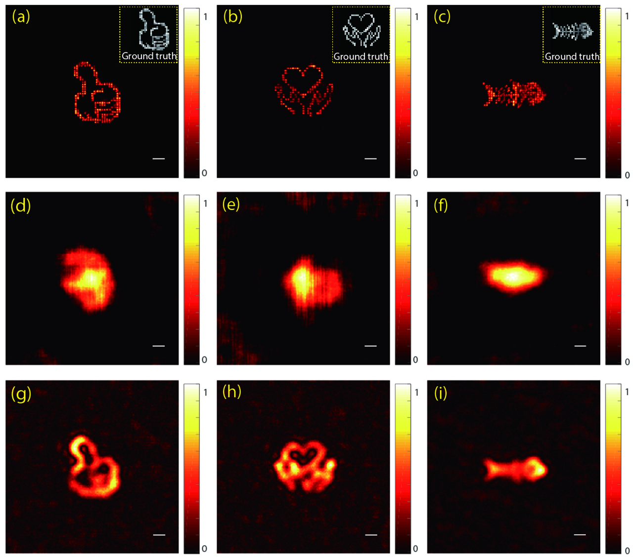
Called stochastic optical scattering localisation imaging, the technique uses an algorithm developed by the team that reconstructs images behind translucent objects, which are objects that light can pass through partially.
Photos of the object are taken with a normal digital camera, with a light source behind the object. The light diffusion patterns in the photos are then studied by the algorithm.
In their experiments, the researchers were able to reconstruct images under egg shell membranes, using a laser as a light source.
This is the first time it has been proven possible to reconstruct images behind translucent objects using a non-invasive technique, the team believes.
The team said this technique has promising applications for life sciences, as examples of images that could be reconstructed in the future include those of cells and viruses that lie just beneath the skin.
Assistant Professor Steve Cuong Dang, from NTU's School of Electrical and Electronic Engineering, who led the study, told The Straits Times: "Our imaging technique demonstrates that we can now not only see through translucent media, but also visualise nanometer-scale objects at unprecedented levels of detail. It can be beneficial to scientists in the life sciences who wish to study bacteria, viruses and components within a cell."
This technique can be used to study objects as small as 100 nanometres, making it comparable to, or even better than, ultra-high resolution microscopes that are currently used to study objects of this size, the team said.
The team hopes that this technique can be a much more affordable alternative to ultra-high resolution microscopes, which range from hundreds of thousands to millions of dollars.
Another advantage of this technique lies in its non-invasive nature.
"We can image through skins and cell walls, without killing the cells or making the skin optically transparent," Prof Dang said.
This means that objects like skins and cell walls can be practically invisible with this technique, and this removes the need for contrast agents.
Currently, contrast agents are typically injected into tissue samples to provide contrast between the objects of interest - cancer cells, for instance - and their background, such as healthy skin tissue.
The new method also provides images at a resolution that is eight times the limit of an optical microscope, which is a key advantage, said Professor Peter Török, Professor of Optical Physics at NTU, who was not involved in the study.
Prof Török, who is also director of imaging at the Singapore Centre for Environmental Life Sciences Engineering, added that other techniques are limited in terms of resolution, speed in processing images, and their need for contrast agents.
The team is currently exploring other sources of light that are more accessible, affordable and practical, while ensuring that the imaging technology can produce the same level of ultra-high resolution images.
The team has filed a patent for this technique, and Prof Dang estimated that it would take around three years for it to be commercialised.
Prof Dang said: "It is our hope that one day, we will be able to take images through the body by light rather than X-ray."