
NEW YORK • The images tell a heartbreaking story: Zika's calamitous attack on the brains of babies - as seen from the inside.
With a macabre catalogue of brain scans and ultrasound pictures, a new study details the devastation done to 45 Brazilian babies whose mothers were infected with Zika during pregnancy.
The study, published on Tuesday in the journal Radiology, is the most comprehensive collection of such images so far, and it reveals a virus that can launch assaults beyond microcephaly, the condition of unusually small heads that has become the sinister signature of Zika.
Most of the babies in the study were born with microcephaly, but many of them also suffered other impairments, including damage to important parts of the brain: the corpus callosum, which connects the two hemispheres of the brain; the cerebellum, which plays a significant role in movement, balance and speech; and the basal ganglia, which are involved in thinking and emotion.
"It's not just the small brain, it's that there's a lot more damage," said Dr Deborah Levine, an author of the study and a professor of radiology at Harvard Medical School in Boston. "The abnormalities that we see in the brain suggest a very early disruption of the brain development process."
The findings also raised concerns about whether babies born without such obvious impairments could develop brain damage as they grow up. For example, almost all the babies in the study had problems in the cortex, including clumps of calcium and neurons that did not reach the right location in the brain.
Because the cortex keeps developing after birth, Dr Levine said, "we're concerned that there might be mild cases that we haven't seen yet, and we should keep monitoring the babies after birth to see if they have cortical abnormalities".
The images studied came from 17 babies whose mothers had a confirmed Zika infection during pregnancy and from 28 without laboratory proof but with all indications of Zika.
Dr Levine described Zika's three-pronged attack. The virus causes dysgenesis, in which parts of the brain do not form normally.
It causes obstruction, primarily because it keeps the ventricles or cavities of the brain so full of fluid that they "blow up like a balloon", she said.
And it destroys parts of the brain after they form. "The brain that should be there," Dr Levine said, "is not there."
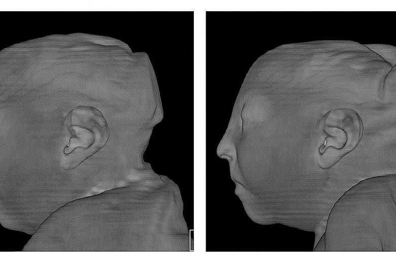
The images in the study include scans of twin girls, who both developed microcephaly. The pictures show folds of overlapping skin and a sloping forehead, indications not only that the brain is smaller, but also that the forebrain has not developed normally.
The brain scans show that the babies have little or no corpus callosum to help one side of the brain communicate with the other.
"The corpus callosum is very important," Dr Levine said. "It is the largest structure that connects the two sides of the brain."
Abnormalities in the corpus callosum were found in 38 babies.
"In some of them, I'm sure it wasn't there," she said. "That either means it never formed or it was destroyed."
NEW YORK TIMES