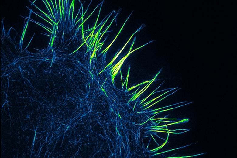
Beautiful, yet deadly, a cancer cell probes its environment with spiky protrusions called filopodia in this award-winning picture.
Scientists at the Agency for Science, Technology and Research's Institute of Medical Biology used a state-of-the-art super-resolution imaging technique known as structured illumination microscopy to illustrate how fine, delicate and complex these cellular structures are.
Captured with a resolution of 120nm (a nanometre is a billionth of a metre), the image shows a single neuroblastoma cell induced by biochemical signals to form filopodia protrusions.
Neuroblastoma is a type of cancer that forms in certain types of nerve tissue.
This photo recently won first prize in Cell-ebrate Science, an imaging competition open to entries from South-east Asia and Taiwan.