Understanding how cells change shape has opened up new avenues for biomedical research, particularly in tissue engineering, which is providing new treatment options for the repair of damaged tissues or organs.
Research on fruit fly embryos, led by Associate Professor Yusuke Toyama from the Mechanobiology Institute at the National University of Singapore, shows that a cell changes shape through the activity of contractile networks within the cell and neighbouring cells.
His discovery offers insight into how scientists can control a cell's shape.
One possible technique is optogenetics, where light is used to control cellular activities.
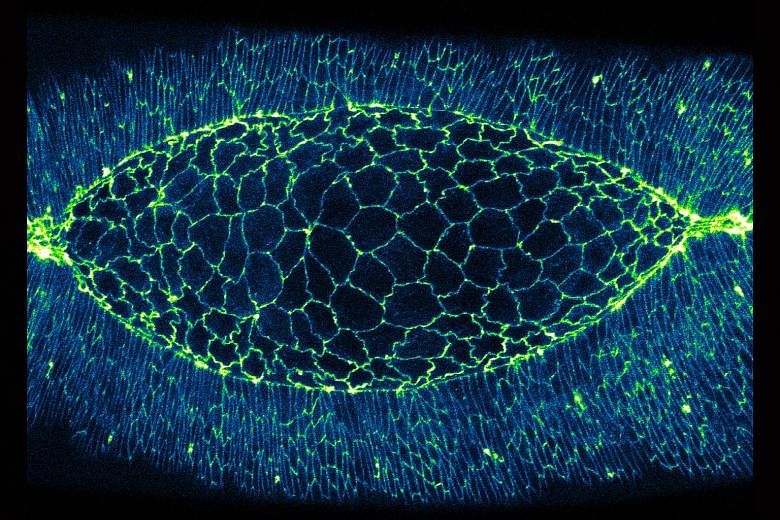
A fruit fly embryo undergoes dorsal closure, where polygon-shaped cells change shape as they "zip up" to seal the embryo.
This makes the cells ideal for studying cell boundary elongation and contraction.
The boundaries are labelled with a fluorescent protein.
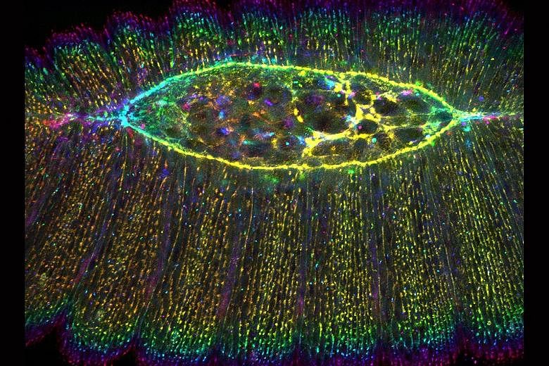
An image of contractile network protein cables, which drive dorsal closure.