
Trying to get a good look at living mouse embryos can be frustrating.
Zoom in on them under strong light and they stop growing; peer at them under a regular microscope and their movements look flat and unnatural.
But Dr Nicolas Plachta of the Institute of Molecular and Cell Biology at the Agency for Science, Technology and Research (A*Star) has found a way to scan these embryos with laser microscopes to get a 3D movie of how they grow and change.
This helps researchers like him better understand how cells develop, and could even pave the way for more effective in-vitro fertilisation (IVF) in the future.
"During the very early stages of development, the embryo of a mouse is identical to that of a human," explained Dr Plachta, who is a senior principal investigator.
"They look amazingly similar."
Traditionally, scientists carrying out IVF can weed out only those embryos that show obvious signs that they are not developing well.
The rest tend to look much the same.
"The big problem for clinicians is: which one should I implant in the uterus of the woman?" Dr Plachta said.
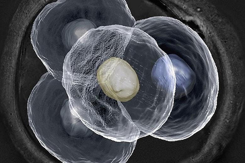
Using the $1.4 million laser scanning microscope, Dr Plachta can see inside each individual cell from all directions, at different points in time.
This means that he can better track what is going on inside the embryo without disrupting its development.
Just like the magnetic resonance imaging (MRI) scans done in hospitals, the laser scanning technique takes cross sections of each embryo and reconstructs them in 3D for scientists to inspect virtually.
"But with an MRI, you see things with very low resolution. You don't even detect single cells," Dr Plachta said.
For example, it was once thought that all cells in an embryo are the same and start becoming differentiated from each other on a random basis.
But using the laser scanning technique, Dr Plachta and his team found that certain proteins, called transcription factors, influence this process.
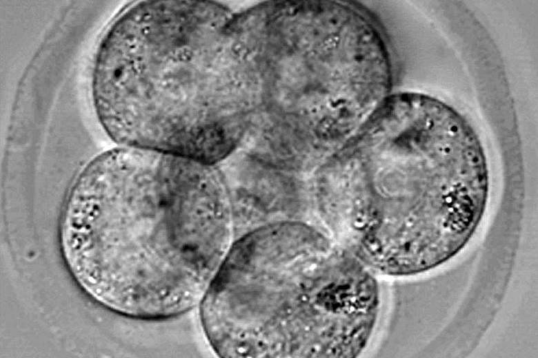
In fact, cells can start becoming differentiated from very early on, when the embryo only has four cells.
Their findings were published in the prestigious international journal Cell earlier this year.
While the laser scanning technique has been used to study the embryos of zebrafish and fruit flies, those of mammals tend not to do as well, said Dr Plachta.
Only in recent years has technology improved such that mammal embryos can be scanned without damaging them.
And knowing how they develop is the first step to fixing problems when they crop up, such as during IVF cycles.
Said Dr Plachta: "To understand what went wrong, you need to know how things are put together in the first place."